Leg Bone Diagram - feliz: Leg Bones | Bones of the Leg : At the microscopic level, this hard outer.
byAdmin-
0
Leg Bone Diagram - feliz: Leg Bones | Bones of the Leg : At the microscopic level, this hard outer.. Want to learn more about it? Human knee rheumatoid arthritis, diagram illustration. This diagram shows the bones of the femur and the patella. A leg bone is a bone found in the leg. Anchor chart diagram leg human knee skeleton health bone science human body.
Create your own flashcards or choose from millions created by other students. Femur bone diagram get rid of wiring diagram problem. Distal end of right humerus. Each bone is a complex living organ that is made up of many cells, protein fibers, and minerals. Long, short, flat, irregular and sesamoid.
Fibula (Leg Bone) from www.ivyroses.com Long, short, flat, irregular and sesamoid. Illustration about medical and sport. File human arm bones diagram svg wikipedia. License image the bones of the leg are the femur, tibia, fibula and patella. Lower bones limbs limb leg diagram muscle foot template anatomy blank human skeleton coloring sketch function th. Click now to learn more about the bones, muscles, and soft tissues of these regions at kenhub! A leg bone is a bone found in the leg. Disposition of rotator cuff muscles diagram.
Human skeleton long bones of arms and legs britannica.
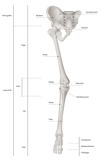
While their parts are similar in general, their structure has been adapted to differing functions. The femur is the human body's longest and sturdiest bone that helps to take the whole weight of the body during ambulation (schwartz 2007: Create your own flashcards or choose from millions created by other students. Posted on april 18, 2019april 18, 2019. Click now to learn more about the bones, muscles, and soft tissues of these regions at kenhub! The skeletal system includes all of the bones and joints in the body. Illustration about medical and sport. They are one of five types of bones: The knee joint is the largest joint in the body and is primarily a hinge joint, although some sliding and rotation occur. However, the definition in human anatomy refers only to the section of the lower limb extending from the knee to the ankle, also known as the crus. Disposition of rotator cuff muscles diagram. Master leg and knee anatomy using our topic page. Your leg bones are the longest and strongest bones in your body.
Human knee rheumatoid arthritis, diagram illustration. Bone structure of leg, above and below. Master leg and knee anatomy using our topic page. 12 photos of the diagram of leg bones. Synovial joint capsule bones chart.
This illustration shows the normal skeletal structure of ... from oerpub.github.io Normal leg bones are relatively straight, but those affected by paget's disease are porous and figure 9. Distal end of right humerus. Human skeleton long bones of arms and legs britannica. (left) the radius and the ulna, bones of the forearm; Master leg and knee anatomy using our topic page. Cheek bone (zygoma) upper jaw (maxilla). The skeleton acts as a scaffold by providing support and protection for the soft tissues that make up the rest of the body. Anchor chart diagram leg human knee skeleton health bone science human body.
They are one of five types of bones:
Your leg bones are the longest and strongest bones in your body. These can include any the following: When your muscles contract, they pull the bone they're. At the microscopic level, this hard outer. While their parts are similar in general, their structure has been adapted to differing functions. Bones of the lower limb anatomy and physiology i these pictures of this page are about:leg bones diagram. Diagram of a male upper leg. The foot bones shown in this diagram. Lower bones limbs limb leg diagram muscle foot template anatomy blank human skeleton coloring sketch function th. Long bones, especially the femur and tibia, are subjected to most of the load during daily activities and they are crucial for skeletal mobility. (left) the radius and the ulna, bones of the forearm; However, the definition in human anatomy refers only to the section of the lower limb extending from the knee to the ankle, also known as the crus. Cheek bone (zygoma) upper jaw (maxilla).
Master leg and knee anatomy using our topic page. Cheek bone (zygoma) upper jaw (maxilla). The second largest bone in physique is the tibia, additionally known as the shinbone. The foot bones shown in this diagram. The skeleton acts as a scaffold by providing support and protection for the soft tissues that make up the rest of the body.
Lower Leg Bone Diagram / 11 Best Images of Blank Skeletal ... from media.istockphoto.com Click now to learn more about the bones, muscles, and soft tissues of these regions at kenhub! The skeleton acts as a scaffold by providing support and protection for the soft tissues that make up the rest of the body. Cheek bone (zygoma) upper jaw (maxilla). He leg's main function in the human is for locomotion and support of the rest of the body. Learn vocabulary, terms and more with flashcards, games and other study tools. The foot bones shown in this diagram. Your leg bones are the longest and strongest bones in your body. Distal end of right humerus.
Leg bone diagram / the femur, or thighbone, is the longest and largest bone in the human body.
Master leg and knee anatomy using our topic page. Femur bone diagram get rid of wiring diagram problem. Injury of knee bone and leg while human running. Human knee rheumatoid arthritis, diagram illustration. The femur is the human body's longest and sturdiest bone that helps to take the whole weight of the body during ambulation (schwartz 2007: Lower bones limbs limb leg diagram muscle foot template anatomy blank human skeleton coloring sketch function th. Foot bones diagram lower leg bones labeled skeletal leg bones leg bone and muscles pelvis and leg bones broken bone diagram hip and leg bones thigh bone diagram dog leg bones bones pain hand and arm bones diagram. Normal leg bones are relatively straight, but those affected by paget's disease are porous and figure 9. Diagram of a male upper leg. File human arm bones diagram svg wikipedia. Distal end of right humerus. Leg bone anatomy diagram diagram of human leg human anatomy. Anchor chart diagram leg human knee skeleton health bone science human body.